
NEWS
The Basal Ganglia Atlas Shows Striatum Zones Fading With Age
A 25.5 million-cell basal ganglia atlas finds six striatal zones that blur with age, tying Parkinson’s and Huntington’s to fading cell identity.

Researchers released a basal ganglia atlas of 25.5 million cells on November 12, 2026, and found six striatal zones that blur with age.
The map covers humans, macaques, and marmosets. It is the first major product of the NIH BRAIN Initiative Cell Atlas Network, a $500 million effort to catalog brain cell types. In the striatum, the largest piece of this circuit, six molecular zones lose their borders as people age, including in tissue hit by Parkinson’s and Huntington’s disease.
Hidden Histology in the Human Striatum
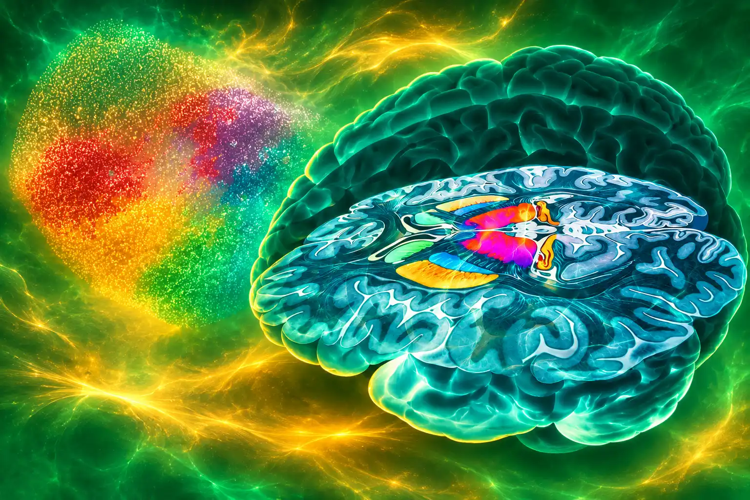
Unlike the cortex, the striatum does not come with neat lines you can see under a microscope. Broad Institute labs used Slide-tags, a method that pins a location to each cell’s gene reads, and surveyed 1.1 million cells across the striatum of 19 postmortem brains. They found six new molecular zones in the striatum, each with its own mix of projection neurons and support cells, and each tied to a different set of brain jobs.
We call it hidden histology because the cells in these zones all look the same under the microscope, but have very strong transcriptional differences and are strikingly consistent from person to person.
Evan Macosko, core member, Broad Institute of MIT and Harvard
Macosko, who also holds appointments at Massachusetts General Hospital and Harvard Medical School, said those “zip codes” showed up in macaques as well. In mice, the same six domains line up with wiring, which is a hint they do real work, not just paint pretty maps. The team then took the zone labels into more than 130 brains from a companion study and watched the borders go soft. Gene-activity gaps between zones shrank with age, and the fade was stronger in regions damaged by Huntington’s and Parkinson’s disease.
That is a different picture from the usual story of those illnesses, which is that particular cells die. Here the cells are still present, and the molecular address that made one patch of striatum unlike the patch next door is what thins out.
Broad scientists mapped six new zones in the brain’s striatum, showed how their boundaries blur with age, and laid groundwork for studying brain disorders and building a whole human brain atlas. https://t.co/EKaI0EvUy1 pic.twitter.com/TZtEK5wX15
— Broad Institute (@broadinstitute) November 12, 2026
Gene Activity Predicts Age to About 5 Years
The same Broad group, with Steve McCarroll’s lab, ran single-nucleus RNA sequencing on 4 million cells from the striatum and the prefrontal cortex of nearly 200 adult postmortem brains. Cell-type balance was steady inside one person. Between people, it swung, especially for non-neuronal cells such as astrocytes and microglia. That is a problem for any “the human striatum” reference built on a handful of donors, and it is why the variation paper exists beside the atlas.
Age moved every cell class they tracked. Projection neurons, the cells that talk to distant brain areas, showed some of the largest gene-activity shifts. University of Pennsylvania neuroscientist Michael Platt, writing about a related BICAN analysis, said that in most cell types with a sex bias, female brains looked molecularly older than male brains of the same age, a pattern that tracks women’s higher Alzheimer’s risk. Spiny projection neurons in the basal ganglia went the other way. Those cells aged faster in male brains, which Platt said may help explain why Parkinson’s disease is more common in men.
THE AGING SIGNAL IN STRIATAL CELLS
- Age clock: Gene activity in any striatal cell type predicts a person’s age to about 5 years, according to the BICAN consortium summary of 150+ donors.
- Support-cell drop: Oligodendrocyte progenitor cells, which mature into the cells that wrap nerve fibers, declined by about 40 percent from age 30 to 80.
- Zone fade: The six striatal zone borders were re-checked in more than 130 brains and grew less distinct with age.
- Person-to-person spread: Glial mix varied more between donors than neuron mix did, so a single reference brain is a poor stand-in for the next one.
Chongyuan Luo, an associate professor at UCLA, reported a developmental rhyme for that adult fade. Spiny projection neurons, also called medium spiny neurons, mature in a tight, synchronized wave, unlike cortical cells, which come of age on staggered clocks. Striosomal cells in that class even show FOS and JUN activity, a mark of firing, as early as mid-gestation. A population that is born in lockstep can also lose its regional accent in lockstep decades later.
Why the Basal Ganglia Came First
Hongkui Zeng, executive vice president and director of Brain Science at the Allen Institute, said disease is always cell-type-specific, so a usable parts list has to come before any serious attempt to fix a circuit. The basal ganglia sit deep in the brain and handle movement, habit, decision, and mood. When they fail, the diagnoses look unrelated at the clinic, and they share this hardware.
THE ROAD TO THIS ATLAS
- 2014: The NIH BRAIN Initiative begins as a national project to study brain cells and circuits.
- 2022: BICAN launches with 11 grants projected at $100 million a year for five years, a $500 million program.
- November 12, 2026: The basal ganglia package of 26 studies in Cell Press journals is published, spanning Cell, Cell Reports, Cell Reports Methods, Cell Systems, Cell Genomics, and Neuron.
- December 1, 2026: BICAN hosts a public webinar on cell types in the map, from 9:00 to 10:30 a.m. PT.
Ana Muñoz-Manchado, a neuroscientist at the University of Cádiz who studies this circuit in degenerative disease and was not on the core atlas team, called it “a massive dictionary that we needed in the field.” Macosko put it in plainer American: this region is “grand central” for disease in both psychiatry and neurodegeneration.
THE CONDITIONS THIS CIRCUIT ALREADY TOUCHES
- Parkinson’s and Huntington’s: Movement circuits in the striatum are the classic injury sites, and the new zone map lands on those same patches.
- Schizophrenia and bipolar disorder: Genetic risk now traces to a rare D2 neuron that makes a serotonin receptor, named in the Allen package.
- Autism: A University of California, San Francisco study in the set reports gene changes in specific basal ganglia cell types.
- Addiction, Tourette syndrome, and OCD: Habit and reward loops run through the same nuclei the atlas now names cell by cell.
- Alzheimer’s in the caudate: BICAN lists a caudate application among the first disease uses of the map.
Comparing humans, macaques, and marmosets, the teams named 61 cell types conserved across about 76 million years of primate evolution. Major neuron classes line up. Primates also show expansions and extra interneuron specialty, with species-specific gene-control tweaks. That is the practical reason the mouse, already mapped in earlier BICAN work, is in the stack: it is the lab stand-in, and it is no longer an unaligned one.
25.5 Million Cells From Six Human Brains
The headline cell count is real, and so is the small number of people behind the core census. The cross-species atlas profiled 21.0 million human cells from six brains, 3.0 million cells from three rhesus and cynomolgus macaques, and 1.5 million cells from two marmosets. Hundreds of scientists at 55 institutions in nine countries, more than 600 consortium members, did the work.
THE CORE CELL CENSUS
| Species | Cells profiled | Donors in the core census |
|---|---|---|
| Human | 21.0 million | 6 |
| Rhesus and cynomolgus macaque | 3.0 million | 3 |
| Marmoset | 1.5 million | 2 |
| Total | 25.5 million | 11 |
Those six human brains are not the nearly 200 donors in the Broad variation study, and they are not the 19 brains used to draw the six zones. Mixing those counts would make the census look broader than it is. Glial mix already differs sharply from person to person in the larger sample. A six-brain “human” reference is a starting scaffold, which is how Allen Institute bioinformatician Michael Hawrylycz described it, not a finished account of how every adult striatum is built.
The measurements go past a gene-activity roll call. Teams added DNA marks that turn genes on and off, how DNA is folded inside the nucleus, electrical recordings, and 3D location. Ed Lein, executive vice president and director of Brain Health at the Allen Institute, said the molecular classes agreed across those data types and across labs, which is the test that this is organization in the tissue rather than a clustering artifact. Joseph Ecker, a professor at the Salk Institute and a corresponding author on the framework paper, said the first step had to set the standard, because the end goal is a community reference for later neuroscience, not a one-off catalog.
A Rare Neuron and a Narrower Parkinson’s Therapy
Nelson Johansen, an assistant investigator at the Allen Institute, and colleagues described a neuron that older striatum catalogs missed. Its working name is STR D2 SMYD2-HTR7: a D2 spiny projection neuron that expresses SMYD2 and the serotonin receptor HTR7. The cell is rare, and it carries extra genetic risk for schizophrenia and bipolar disorder.
Schizophrenia has been associated with the striatum, but it has been difficult to define which neurons are vulnerable. Our study revealed a rare neuron type expressing the serotonin receptor HTR7 that was not found in previous studies, which carries elevated schizophrenia risk. This atlas narrows the genetic disease signal down to a specific population that researchers can now explore.
Nelson Johansen, assistant investigator, Allen Institute
The same Allen rare neuron type expressing the serotonin receptor package reports a second, more applied result. Scientists built a smarter version of an existing Parkinson’s gene therapy aimed at a subset of basal ganglia cells. In animal models it cut the tremor that defines the disease, and the teams said the targeted shot was both more effective and less invasive than the broader methods now used in people, which makes it easier to scale. That is still a model result. It is also the first time this atlas has been used as a targeting menu rather than a poster.
Zeng’s line sits under both findings. If a disease lives in one cell class, a therapy that hits every cell in the neighborhood is the old design. A map that names the class is how you stop doing that.
Open Data for the Rest of the Brain
Lein said the basal ganglia project was designed as the template for the whole brain, and that the shared cell names are the point. For the first time, every study in the package used the same cellular framework. Labs can now park their own samples on that grid and inherit what is already known about each type. Kiku Ichihara, managing director of the Center for Human Brain Variation at the Broad Institute, said this is the start of the atlas, and that the same tools will now move across the rest of the brain.
The consortium put the open basal ganglia atlas and data tools on a public portal the day the papers went up. Zeng called the effort a social-engineering experiment as much as a biology one: dozens of labs, different methods, one integrated object. John Ngai, director of the NIH BRAIN Initiative, said the atlas is aimed at the cell basis of neurologic, psychiatric, and substance-use disorders, which is the job the 2022 awards were written to do.
A public webinar on December 1, 2026, will walk through the cell types. The whole-brain census is still being built. What this package already changes is narrower, and harder to walk back: the striatum has an interior map, that map fades with age, and the fade sits on the same ground Parkinson’s and Huntington’s already occupy.
Frequently Asked Questions
How Can Researchers Access the Basal Ganglia Atlas?
Primary files sit in the NIH Neuroscience Multiomic Archive, and the Allen Institute’s ABC Atlas lets labs browse the single-cell and spatial data. MapMyCells assigns a lab’s own cells to the consensus taxonomy, and the Harmonized Ontology of the Mammalian Brain Anatomy applies the same structure names in human, macaque, and marmoset MRI templates, so a caudate in one species is not a renamed mystery in the next.
What Is Slide-tags and What Did It Add?
Slide-tags is a spatial method invented by Evan Macosko and Broad colleague Fei Chen that puts location coordinates on single-cell gene reads. One published section plot places 92,100 cells on their original spots and colors them by zone, with gray for the surrounding white matter, which is how the six domains became visible without a classic stain.
What Are the Enhancer-AAV Vectors in the Package For?
The BRAIN Armamentarium released more than 400 enhancer-AAV vectors for cell-type-specific targeting, ranked across species and checked against the consensus taxonomy. Labs can use them to switch genes on in chosen basal ganglia cells in model animals instead of flooding a whole nucleus with a virus.
Does the Atlas Include Mouse Data?
Yes. Teams folded in earlier whole-mouse-brain maps so the primate taxonomy can be aligned to the mouse, the main lab model for Parkinson’s and Huntington’s circuits. BICAN lists four species and more than five measurement types, from gene activity and DNA folding to electrical recordings.
What Does the Name STR D2 SMYD2-HTR7 Mean?
The label is a D2 spiny projection neuron that expresses SMYD2 and the serotonin receptor HTR7. That combination was missing from earlier striatum catalogs, which is why Johansen’s group could finally park schizophrenia and bipolar genetic risk on one rare cell class instead of on the striatum as a blur.
Disclaimer: This article is news reporting on published research and is for information only. It is not medical advice, a diagnosis, or a recommendation for any Parkinson’s, Huntington’s, schizophrenia, or other brain-disease treatment. Readers who have symptoms or treatment questions should consult a licensed physician or neurologist before making health decisions. Cell counts, donor numbers, and study findings reflect the papers and lab statements dated November 12, 2026, and later work may change them.

Airbnb Starts House Rules Reviews as Cities Rewrite Contracts

United’s Computers Failed the Day After Kirby Blamed ATC

Tyson Foods Guides Beef Losses Narrower as Chicken Cools

Saudi Arabia’s Leisure Tourism Falters as Pilgrims Still Come

Himesh Patel Confirms the X-Files Writers’ Room Is Open

AI Server Boards Put Everyday Circuit Materials on Allocation

Glacier Collapse Buried Nepal’s Hydropower Workers in Tunnels

Congo’s Bundibugyo Outbreak Outruns the Licensed Ebola Shots

Pennsylvania Measles Deaths Follow a Long Slide in Coverage

CISA’s Known Exploits Still Come From 2007-Era Bugs
-
 NEWS2 months ago
AI Server Boards Put Everyday Circuit Materials on Allocation
-
 NEWS3 months ago
Glacier Collapse Buried Nepal’s Hydropower Workers in Tunnels
-
 NEWS3 months ago
Congo’s Bundibugyo Outbreak Outruns the Licensed Ebola Shots
-
 NEWS3 months ago
Pennsylvania Measles Deaths Follow a Long Slide in Coverage
-
 NEWS3 months ago
CISA’s Known Exploits Still Come From 2007-Era Bugs
-

 NEWS3 months ago
Texas Judge Freezes the SEC Eligibility Ban for 41 Athletes
-

 NEWS3 months ago
Germany Can Miss Its 2026 Climate Cap Without Penalty
-

 NEWS2 months ago
Pig Kidneys Got Two Men to Human Transplants After Failing